What do healthy gums look like?
Healthy gums are light pink, firm, tightly hugging the neck of the teeth, and do not bleed when brushing. This is a benchmark for comparison with the other images.
When gums begin to discolor or change shape, it may be the first stage of inflammation, as described below.
What does gingivitis look like?
Gingivitis is the stage of inflammation confined to the gum tissue. The gums are red, swollen, and bleed easily, without loss of attachment or bone loss due to disease. This condition can be completely reversible if the cause is detected and controlled early. Gingivitis resolves when plaque is effectively removed, usually requiring professional cleaning combined with proper hygiene, and should be evaluated by a doctor rather than waiting for it to heal on its own.
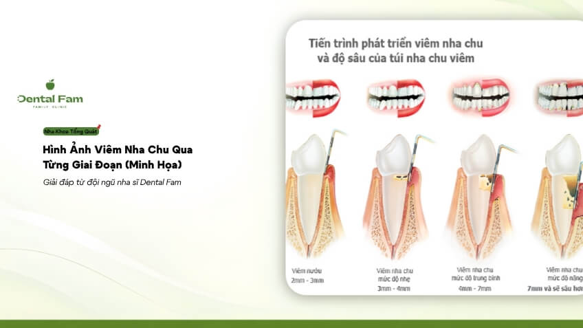
You can compare the differences between gingivitis and periodontitis in more detail. When inflammation spreads to the ligaments and bone, the disease progresses to periodontitis, illustrated in stages below.
Periodontic Disease: Illustrated Stages I to IV
Periodontic disease is divided into 4 stages, from the initial stage (I) to the advanced stage (IV), and the appearance gradually changes from subtle to pronounced. The four images below illustrate the typical signs of each stage; these may vary from person to person, so they are only general illustrations.
Initial Stage
In stage I, the gums may only appear slightly red, slightly swollen, and similar to gingivitis, making it difficult to distinguish with the naked eye. Attachment loss is still minor and can only be detected by a dentist.
Stage 2: More pronounced attachment loss
In stage II, the gums are more noticeably red and swollen, may begin to recede slightly, and the gap between the teeth and gums deepens into periodontal pockets. Periodontal pockets are the spaces between the teeth and gums where plaque easily accumulates and is difficult to reach with a toothbrush.
Stage III: Gum recession and exposed tooth roots
In stage III, gum recession is more pronounced, tooth roots begin to be exposed, teeth feel longer, and may be slightly loose. Significant support tissue has been lost, so the dentist needs to assess the possibility of preserving each tooth.
Stage IV: Gapped and loose teeth
In stage IV, teeth may become increasingly spaced, displaced, and noticeably loose, affecting chewing. Many teeth may have been lost due to periodontal disease, making the case more complex. Gapped and loose teeth don't necessarily mean all teeth need to be extracted: the dentist assesses each tooth based on bone loss and looseness; many cases still have a chance of preservation or restoration after extraction, and no one can promise a guaranteed outcome.
Which image you see that resembles yours isn't enough to draw a conclusion, for reasons explained below.
Why can't images replace a physical examination?
Images only help you compare; only a doctor can determine the true extent of the damage, as most lesions are located beneath the gums and within the bone. Two people with similar-looking gums may still be in different stages, and even without bleeding or pain, they could still be in stage I or II because the damage is often subgingival. Stages usually progress over time, but the rate varies from person to person, related to the grade (level of progression), so you shouldn't wait until your image shows a severe stage before seeing a doctor.
To determine the stage, the doctor will measure the depth of the periodontal pockets, assess attachment loss, check for looseness, and take X-rays. Please refer to the article on signs of periodontitis to recognize the symptoms, and the article on [classification of periodontitis by stage and level of progression] to understand the meaning of each stage. After comparison, you can schedule an appointment at Dental Fam, as described below.
How does Dental Fam help you with your assessment?
Dental Fam assesses the severity using measurements and X-rays, then clearly explains your condition. The doctor will create a personalized treatment plan and choose the appropriate option for each tooth: non-surgical, LASER (laser treatment), or surgical, starting from 500,000 VND per tooth, including examination and consultation.
Periodontal disease images gradually change from swollen and red gums to receding gums, exposed tooth roots, and gaps between teeth, but only an examination can determine the exact severity. Illustrations help you recognize early signs without needing to look at frightening pictures. The main damage is located under the gums and in the bone, so it cannot be assessed visually.
At Dental Fam, the doctor will measure, take X-rays, and advise on treatment options starting from 500,000 VND per tooth, including examination and consultation. Please schedule an appointment to know your exact condition.
Dental Fam - Family Clinic
- Address: NOTT 28, 29, 30, Phuoc Long A Urban Area, Nha Trang, Khanh Hoa
- Hotline: +84 582 582 345 / +84 983 001 768
- Email: info@dentalfam.vn